Robust Theme
Dec 09, 2019 2020-04-08 7:40Robust Theme

Microscopes: Avenues for Success
The opportunities in dentistry to have our field of vision enhanced is nothing new. Surgical loupes have been around since 1876. They were convex lenses that were attached to a headband. We've come a long way. I can remember a few dentists beginning to use them in the 1990’s, mostly the dentists that were involved in learning from places like the L.D. Pankey Institute. Loupes not only help with vision but ergonomically it was a game changer. Magnification can begin at 2.0x and extend all the way to 8.0x, even though I have never seen anything close to that in dentistry. In this day and age most clinicians, dentists and hygienists alike, use loupes in their daily practice.
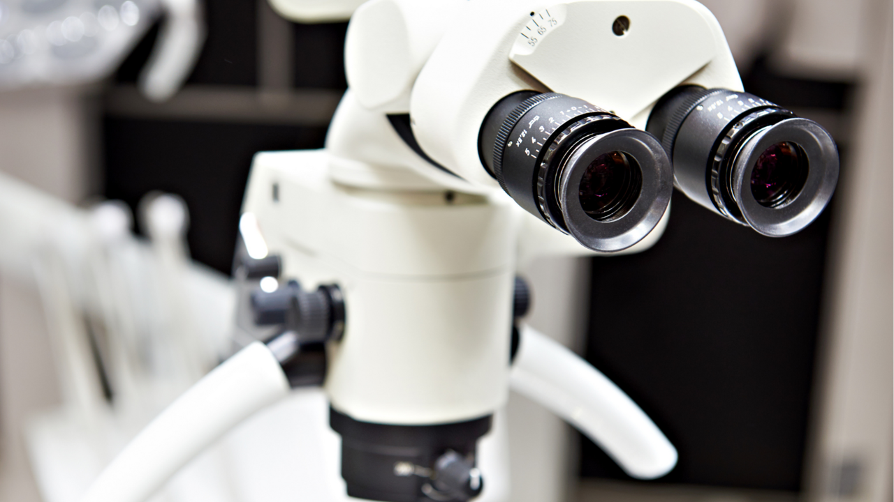
However, the field of vision expanded exponentially with the use of dental operating microscopes. In the 90’s they became a tool for endodontic residencies and in the last 25 years has been adopted for work in all phases of dentistry. The use in periodontal treatment, restorative dentistry and surgery over the last two decades has increased tremendously and the design for viewing during actual clinical work with patients has helped and continue to help treatment outcomes for patients. The microscope gives 400 times more visual accuracy than the naked eye and 100 times the visual information than traditional loops. In a practice setting it can help to locate cracks and fractures, it can facilitate all aspects of endodontic surgery, can help with enhancing photographic documentation and can also reduce the unnecessary destruction of tissue when preparing an access.
Implant practice:
Dental Operating Microscopes
Microsurgery can be a useful asset when placing implants from the soft-tissue procedures, suturing and implant drilling precision. Being able to detect minuscule differences in spacial alignment allows implant osteotomy procedures to be placed precisely between appropriate anatomy and implants. The drilling angle can be positioned relative to landmarks, such as neighboring caps and implant platforms, which allows for precision depth and location. Additionally, small alterations of drill positioning can be detected so corrections to the handpiece can be made, as angular accuracy is of the utmost importance. Only 3-4 mm of the exposed root is needed to calculate angulation between the CEJ and the osseous crest, a measure that would be invisible to the naked eye. Precise angular measurements, drill slant correction, and visual maximization provided by microscopes allow precision placement and optimal positioning for preparing extraction sockets and placing implants during osteotomy microsurgery.
Phase Contrast Microscope
Another microscope that could become an asset to an implant practice is a phase contrast microscope. The ability to evaluate biofilm in a patient's mouth prior to and after an implant is placed gives greater success to the implants long term outcome. The hygienist can collect a subgingival plaque sample and within seconds be able to show a patient various types of bacteria, fungus, parasites, white blood cells and red blood cells that are swimming around their mouths. Seeing what’s on the inside can be a great tool for showing a patient how important home care is. Spirochetes are incredibly active on slides and if they are present in biofilm, bleeding follows because they are extremely inflammatory. The last thing an implant patient needs is inflammation. Whether using a phase contrast microscope alone or together with pathogen testing the benefit to the patient is fantastic.
Dental Endoscopy
Another avenue to consider is a dental endoscope. A miniature fiber-optic camera attached to a probe less than one millimeter in diameter that is then placed below the gumline. The images are magnified 24 to 48 times with LED illumination in the working area and are displayed on a chair side monitor. The field of vision ranges from 3-6mm. The fiber-optic system enlarges the image while also delivering light to the working field which can be critical for successful treatment of periodontitis. Endoscopy has come a long way since the 80’s when the images were not very clear and the education of use was limited. Using an endoscope has enabled clinicians to treat periodontal concerns during initial therapy perhaps making traditional flap surgery unnecessary.
The endoscope can be extremely valuable in implantology allowing visualization of residual cement, microgaps, and overhanging margins that can help prevent peri-implantitis. If during restoration of the implant there was an ability to see residual cement, that could perhaps prevent a future of peri-implantitis. If needing to treat peri-implantitis, using an endoscope would be less traumatic, less costly, less invasive and less morbidity than open-flap surgery. The use can also allow the hygienist to scale and remove contaminants subgingivally more effectively than when doing so with limited visual access. Using the endoscope helps to also prevent over instrumentation which can affect the coating on some implants. As minimally invasive implantologists the advantage of a periodontal endoscopy helps to provide yet another avenue for minimally invasive therapy. As with any new technique there is proper training, practice and patience but the rewards for the dentist and the hygienist are well worth it.
Utilizing technology, especially in areas that lend itself to seeing the working area better will help clinicians to not only execute better treatment for the patient but have better long term success.
