Robust Theme
Dec 09, 2019 2020-04-08 7:40Robust Theme

Biofilm: Dentistry's Nemesis
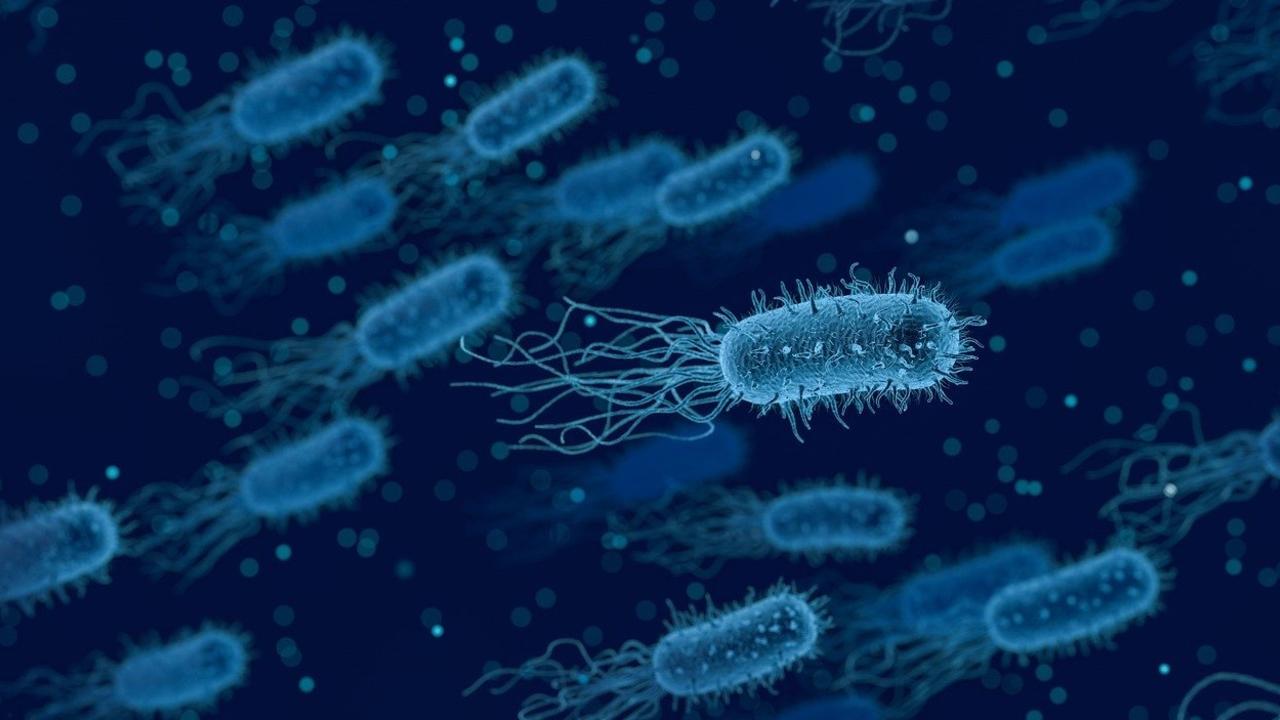
Most bacteria in the body do not exist as a single floating cell. They are in communities called biofilms. To form this biofilm, bacteria first adhere to a surface then generates a polysaccharide matrix that also sequesters calcium, magnesium, iron or whatever minerals are available.
Within the biofilm, one or more types of bacteria share nutrients and DNA that then evade the immune system. It requires less oxygen, fewer nutrients and alters the pH at the core. The biofilm forms a physical barrier that keeps more immune cells from detecting pathogenic bacteria.
Dental plaque has been identified as a biofilm. The nature of biofilm enhances the component of bacteria’s resistance to both the host’s defense system and antimicrobials. If it is not removed regularly the biofilm undergoes maturation, and the resulting pathogenic bacteria complex leads to caries, gingivitis and periodontitis.
Stages of Biofilm Formation
There are 4 stages of biofilm development, initial adherence, lag phase, rapid growth and steady state. The adherence of bacteria to the tooth surface also known as acquired pellicle makes the surface receptive to colonization by specific bacteria. Usually, gram-positive cocci are the first microorganisms to colonize on the teeth. As bacteria shift from planktonic to sessile life, a genetic up-regulation or gene signaling actually promotes the shift. That genetic expression then shifts causing a lag in bacterial growth, but that shift makes it up to 1,000 times more resistant to antibodies, antibiotics and antimicrobial compounds than its single cell state. In the rapid growth stage, the adherent bacteria secrete large amounts of water-insoluble extracellular polysaccharides to form the biofilm matrix. Additional varieties of bacteria adhere to the early colonizers known as coaggregation and the biofilm increases and becomes more complex. This cell division also increases the thickness of the biofilm. The final stage called the steady state phase, the interior of the biofilm slows their growth and become static. In this stage surface, detachment and sloughing occur and some bacteria will travel to form new biofilm colonies.
The Specific Plaque Hypothesis
The specific plaque hypothesis proposes that only certain microorganisms are pathogenic. There are hundreds of species of microorganisms in periodontal pockets but fewer than 20 are routinely found in increased proportions. Specific virulent bacterial species activate the host’s immune and inflammatory responses that then cause bone and soft tissue destruction.
In 1968 Dr. Sigmund Socransky classified the bacterial species involved in the initiation and progression of periodontal disease.1,2 He classified several complexes of bacteria dividing them into groups then labeled them in colors. The categories were based upon the pathogenicity of the bacteria and their role in the development of plaque or biofilm.
His color designations were blue, yellow, green, and purple that are early colonizers of the subgingival flora. The purple and green complexes present in periodontal pockets but are not significantly associated with signs of periodontal disease progression. Orange and red complexes are late colonizers associated with mature subgingival plaque. Orange complex bacteria include: Fusbacterium nucleatum, Prevotella intermedia, Prevotella nigrescens, Peptostreptococcus micros, Streptococcus constellatus, Eubacterium nodatum, Campylobacter showae, Campyllobacter gracilis and Campylobacter rectus. The species in this group are closely associated with each other and species to the red complex.
The bacteria in the red complex are more likely to be associated with clinical indicators of periodontal disease such as periodontal pocketing and clinical attachment loss. Red complex and the individual species in that group are strongly associated with bleeding on probing. Those bacteria are Porphyromonas gingivalis, Tannerella forsythia, and Treponema denticola. They have a strong relationship with pocket depths and P. gingivalis alone or in combination exhibits the deepest mean pocket depths. Since Dr. Socransky’s original research another pathogen has been added to the list of high risk, Aggregatibacter actinomycetemcomitans.
Dental biofilm is a complex, organized microbial community that is the primary etiologic factor for the most frequently occurring oral diseases, dental caries and periodontal disease. Biofilm can’t be eliminated but it can be reduced and controlled. Teeth comprise 20% of the mouths surfaces so using antimicrobial rinses can help to control biofilm that brushing, flossing and irrigators can’t. Biofilm is found in dental unit water lines, on prosthetic appliances, the tongue, biomaterials in dental restorations and on oral mucous membranes. Thorough and diligent oral hygiene practices taught to patients by clinicians and in return how clinicians manage biofilm in the operatories we can make great gains in management.
1. Socransky SS, Haffajee AD, Cugini MA, et al.. Microbial complexes in subgingival plaque. J Clin Periodontol. 1998;25: 134-144.
2. Socransky SS, Haffajee AD. Periodontal microbial etiology. Periodontol 2000. 2005;38: 135-187.
*Please read a companion blog: Guided Biofilm Therapy-A Must for Implant Therapy
